This week I viewed my MicroAquarium™ for the last time. It was relatively less active than the previous viewing but still active enough to spot life almost anywhere. I spotted many of the organisms identified in past blog entries before coming across the protozoa in Figure 1.
Figure 1: Photo of Notosolenus sp. taken during movement.
Note the prominent anterior flagella. (Lee, 1985)
Figure 2: Short animation taken from video of Notosolenus sp.
(Lee, 1985)
Video 1: Digital video of Notosolenus sp.(Lee, 1985)
I eventually identified the organism as the genus Notosolenus. (Lee, 1985)
Some characteristics that helped with identification were the colorless
appearance, egg-shaped body, and the presence of two flagella. Both
flagella were positioned anteriorly on the organism but only one was
clearly visible. The second shorter flagella was dragged alongside its
body providing stabilization. (Lee, 1985) The primary flagella thrashed
in all directions like an out of control fire hose. I first assumed the
round structures inside the body (as seen in Figure 2) were organelles. I
later learned that this genus was colorless and most organelles were
unable to be viewed with a compound light microscope. (Lee, 1985) I
hypothesized that the greenish spots inside of the body were algae
consumed by the organism via phagocytosis. This was a gluttonous protozoa.
I revisited my MicroAquarium™ on 11/06/2013. I was able to spend a great deal of time analyzing my MicroAquarium™ with the compound light microscope and recording my findings with digital video equipment. This was the most "active" viewing period experienced so far. I feel this was due to a combination of the added food source and my growing competence with the microscope and digital recording equipment. I was able to spot life in almost every area of my MicroAquarium™. I was also able to see small organisms darting through the water without the aide of magnification.
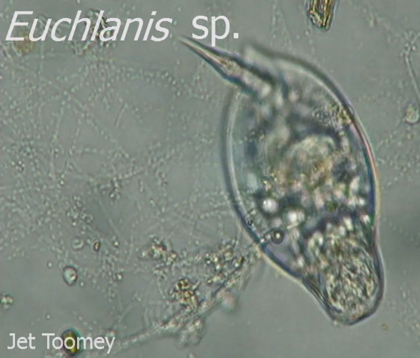
Figure 1: Photoof Euchlanis sp. captured during
movement while feeding in an open area of the
MicroAquarium™ (Pennak, 1953)
I viewed many of these larger organisms and identified them as Euchlanis sp. (Pennak, 1953) After researching this organism I can confidently say that it is the adult form of the unknown organism I documented in Week 2. I learned Euchlanis is the genus for all rotifers. (Pennak, 1953)These organisms appeared more complex than most of their peers. They had two "tails" on their posterior that they often pulled together so that it looked like one large structure. (as seen in Figure 1) Many different organs were visible through their transparent bodies. One feature unique with rotifers are a circle of cilia at their anterior end which create a current that is used to funnel food into their mouths. (Davidson, 1998) Once the food enters their mouth it's crushed by an anatomical device called a mastax which is basically a spiked pharynx from hell. I watched the Euchlanis sp. consume anything within proximity that was able to physically fit into their mouths. (as seen in Video 2) They appeared in greater numbers around vegetation such as Fontinalis sp. (Mcfarland, 2013) The appetite of the Euchlanis sp. is insatiable; I found one with both of its "tails" wedged tightly in branching vegetation and it continued to eat everything around it while being minimally phased by its presumable death sentence. (as seen in Video 3)
Video 1: Footageof Euchlanis sp. consuming debris and
moving rapidly in an open area of the MicroAquarium™
(Pennak, 1953)
Video 2: Footageof Euchlanis sp. feeding on and around
the vegetation Fontinalis sp. (Pennak, 1953)
Video 3: Footageof Euchlanis sp. stuck on the vegetation
Fontinalis sp. but still taking time to consume particles
and debris within its proximity. (Pennak, 1953)
Another interesting organism I found and later identified was theDifflugia sp. (Patterson, 1996)This organism belongs to a major group of amoeboid protozoa in the kingdom protista. Most of the time these organisms are found to be heterotrophs and use pseudopods to move around and capture prey.
Figure 2: PhotoofDifflugia sp.(Patterson, 1996) Note the
silica like outer shell and irregular shape.
If you look closely at the included footage you can see the near transparent pseudopods used to push and pull the organism in different directions. What makes this amoeboid so interesting (in my humble opinion) is how it builds its exterior shell. When I first spotted this organism I honestly thought it was some bizarre or damaged diatom. This was due to what looked like a hard silica shell-like outer layer. It wasn't organized and symmetrical like the diatoms I'd previously documented, instead the outer shell was chaotic and random. I learned that this outer shell is created by the Difflugia sp. using particles gained from its environment or prey. (Patterson, 1996) For example, they consume diatoms and use the silica particles for their shell. This type of shell is referred to as being agglutinate. Amazingly, many Difflugia select particles of certain size and shape and construct a species specific shell. That blows my mind.
Video 4: FootageofDifflugia sp. using pseudopods to
grab and pull itself to a piece of debris. (Patterson, 1996)
Video 5: Additional footageofDifflugia sp. using
pseudopods near debris. (Patterson, 1996)
That's all for now. I will be spending more time observing my MicroAquarium™ this week, so stay tuned for another update. I recently checked my hit counter for this blog and was extremely disappointed. I was expecting hundreds of thousands, if not millions of unique visitors per day and the reality of the data hit me like a ton of bricks; seven unique visitors and 120+ total hits. After days of deep depression I've come to terms with my popularity (or lack thereof) and decided that blog views are not about quantity but quality. Who needs millions of views from ordinary people when I'm graced with the quality blog hits of exceptional people like you?
I spent some time viewing my MicroAquarium™ on 10/30/2013. My lab instructor added one beta food pellet on to my MicroAquarium™ on 10/25/2013. (McFarland, 2013) The following is detailed information on the food source added: "Atison's Betta Food" made by Ocean Nutrition, Aqua Pet Americas, 3528
West 500 South, Salt Lake City, UT 84104. Ingredients: Fish meal, wheat
flower, soy meal, krill meal, minerals, vitamins and preservatives.
Analysis: Crude Protein 36%; Crude fat 4.5%; Crude Fiber 3.5%; Moisture
8% and Ash 15%.
(McFarland, 2013) I noticed more activity since the last viewing; the majority of which appeared to be new organisms. The beta food pellet had disintegrated a great deal and was spread thinly around the left side of the MicroAquarium™. Organism activity was greatest around this area. I was unable to locate the diatom Surirella oblonga that was identified the previous week. (Prescott, 1954)
However, it was difficult to navigate anywhere inside the MicroAquarium™ without spotting a quick moving circular organism that I eventually identified as Anisonema sp. (Patterson, 1996) This organism could be found almost everywhere inside the aquarium but was concentrated in the area of the food pellet. (as seen in Figure 1) This organism didn't have any visible orifice and I didn't observe it eat. However, it was found in higher volume around the beta pellet and plant life. They moved in a very "choppy" or "clumsy" fashion. I focused on watching their movement and one of things I noticed was their ability to "pop" into different directions, including reverse, at any given time. (as seen in Video 1 and Video 2) This odd movement helped identify the organism. The identification guide stated that Anisonema sp. can be distinguished by their jerky movement and ability to contract backwards. (Patterson, 1996)
Figure 1: Digital photograph of Anisonema sp. (Patterson, 1996)
Video 1: Digital video ofsingle Anisonema sp.(Patterson, 1996)
Video 2: Digital video of multipleAnisonema sp. (Patterson, 1996)
I learned that this unique movement was due to the flagella used for movement. A smaller and twisted anterior flagella flails around like a crazy propeller while a larger recurrent flagella stabilizes the organism and helps steer it. This organism was multicellular, motile, and displayed bilateral symmetry.
On 10/23/2013 I returned to the lab and spent over two hours viewing my creation. I started by briefly viewing my MicroAquarium™ with the compound light microscope. This allowed me to compare my findings with the previous session in which I used only a compound light microscope. I quickly realized it was much more difficult to find a noticeable living organism. It seemed like everything "settled" over the course of the previous week. I noticed movement near and under the surface of the debris on the bottom. I then moved to a compound microscope with digital video recording capabilities.
This microscope was able to produce a more clear and vibrant viewing experience than the previous compound microscope. However, there was a learning curve. The digital setup in lab requires the user to locate an organism via live image on a monitor and then switch the source of the monitor to a PC so that the user may gain control of the recording software and engage the recording process. During the few seconds of this transition a fast moving organism will have moved out of the microscopes field of view. THIS. WAS. FRUSTRATING. I was able to capture one of the fast moving organisms as seen in Figure 2 but was unable to identify it; I will work on this and report back next week.
A great deal of my time was spent positively identifying the organism Surirella oblonga as seen in Figure 1. I found him after slowly scanning the MicroAquarium™ from left to right. As you will see in Video 1, this organism moved slowly and smoothly. This organism appeared to be gliding along with the slight current of the water, but this was disproved when it stopped completely at times and even changed direction. I hypothesized that the organism was taking water in one orifice and expelling in out of another to create propulsion. After researching diatoms and their movement I have discovered my hypothesis was likely incorrect. This diatom probably moves with tiny flagella that are unable to be captured with the equipment at hand. Notice the "cigar" shape and bilateral symmetry.
Figure 1: Digital photograph of Surirella oblonga. (Prescott, 1954)
Video 1: Digital video of Surirella oblonga. (Prescott, 1954)
I went through many books before I was able to positively identify this diatom. I was able to positively identify the organism as Surirella oblonga Ehrenberg with the book "How to Know the Freshwater Algae". (Prescott, 1954) I narrowed the search first visually then by more specific detail. I found a few diatoms with the cigar shape of mine but only one that fit the description perfect. The dead giveaway was the unique transparent linear area running through the center of this organism. This diatom is a non-green algae.
Here is a photo and brief video of the unidentified multi-cellular organism that I captured. There were many of these in my MicroAquarium™ but they were all very active. They also appear to dislike the light output from the microscope. This could be why I found these organisms near the debris in the bottom of the MicroAquarium™.
Figure 2: Digital photograph of unidentified organism.
On 10/16/2013 I created my own MicroAquarium™. I started with a clean
MicroAquarium™ and first applied a series of identification tags to the upper
right corner. These identifications will allow me to distinguish my
MicroAquarium™ from the plethora of others that surround it. I was allowed to
choose from 12 different water sources collected by our lab professor. I selected
water source sample #7. The details of this water sample are listed below: Third Creek at Tyson Park. Knox Co. Knoxville TN Partial shade exposure. N30
57 13.53 W83 56 32.37 824 ft. 10/14/2013 (Mcfarland, 2013)
I used a pipette to suction samples from the bottom, middle, and surface
of water source sample #7. I then carefully added this suctioned water to my
MicroAquarium™ until it was about 1/3 inch from the top. One settled, my
MicroAquarium™ had a few cm of debris on the bottom and the remaining water
above was relatively clear.
I then added two mosses and one plant. The details of each are listed below: Amblestegium
varium(Hedwig) Lindberg. Moss. Collection from: Natural
spring. at Carters Mill Park, Carter Mill Road, Knox Co. TN. Partial shade
exposure. N36 01.168 W83 42.832. 10/13/2013 (Mcfarland, 2013)
Fontinalis sp. Moss. Collected from: Holston
River along John Sevier Hwy under I 40 Bridge Partial shade exposure Holston
River water Shed N36 00.527 W83 49.549 823 ft 10/13/2013 (Mcfarland, 2013) Utricularia gibba L. Flowering plant. A carnivous plant. Original
material from south shore of Spain Lake (N 35o55 12.35" W088o20' 47.00),
Camp Bella Air Rd. East of Sparta Tn. in White Co. and grown in water tanks
outside of greenhouse at Hesler Biology Building. The University of Tennessee.
Knox Co. Knoxville TN. 10/13/2013 (Mcfarland, 2013)
Small portions of vegetation were clipped and then directly submerged into the
MicroAquarium™.
I immediately placed the MicroAquarium™ onto the stage of a compound microscope
and searched my new ecosystem at 40x. I found multiple organisms of similar
species. They had one "eye" centered on their head and used a
tail-like structure to move at a surprisingly quick pace. They had four
"tentacle" structures protruding from around their heads. I'm not
sure if these help with movement or assisted with sensory or eating. They had a
vibrant purple core that was visible through their bodies. They appeared to be
feeding on the debris at the bottom of the tank. As they consumed particles
around them you could see small dots of purple travel from their mouth area to
the centralized purple core visible in their bodies. It was crazy.
I will continue to monitor my MicroAquarium™
on a weekly basis. My findings will be posted to this blog along with photos
and possibly videos. I feel more and more powerful as the days go by.
McFarland, Kenneth [Internet] Botany 111 Fall 2013. [cited Oct 28 2013]. Available from http://botany1112013.blogspot.com/ Davidson M. 1998. Euchlanis Rotifer [Internet]. Tallahassee(FL):Florida State University; [2003 Aug 01, cited 2013 Nov 08] . Available from: http://micro.magnet.fsu.edu/primer/techniques/dic/dicgallery/euchlanissmall.html Prescott G.W. 1954. How to Know the Freshwater Algae. 1st ed. Dubuque (IA): WM. C. Brown Company 211 p. Patterson D. 1996. Free-living Freshwater Protozoa: A Colour Guide. New York (NY): Wiley 223 p. Pennak, Robert W. 1953. Fresh Water Invertebrates of the United States. New York (NY): Ronald Press Co 769 p.
Lee, J. 1985. Illustrated Guide to the Protozoa. Lawrence (KS): Society of Protozoologists 629 p.


